

Hospitals
Understanding the C-Arm Machine: A Clinical and Operational Overview
An expert guide for clinicians and procurement heads on the c-arm machine. Explore its clinical applications, key components, evaluation criteria, and operational best practices.


For any modern operation theatre, cath lab, or interventional suite, the C-arm machine is an indispensable asset. Its ability to provide real-time, intraoperative imaging—fluoroscopy—has fundamentally changed how we approach a vast range of surgical and interventional procedures. It provides the visual guidance necessary for precise instrumentation, implant placement, and diagnostic assessments, directly impacting procedural success and patient outcomes.
This is not just another piece of equipment; it's a dynamic imaging tool that must align perfectly with your facility's clinical demands, workflow, and safety protocols. Understanding its core technology, evaluation parameters, and operational mandates is crucial for any department head or procurement manager tasked with its selection and oversight.
Core Components and How They Function
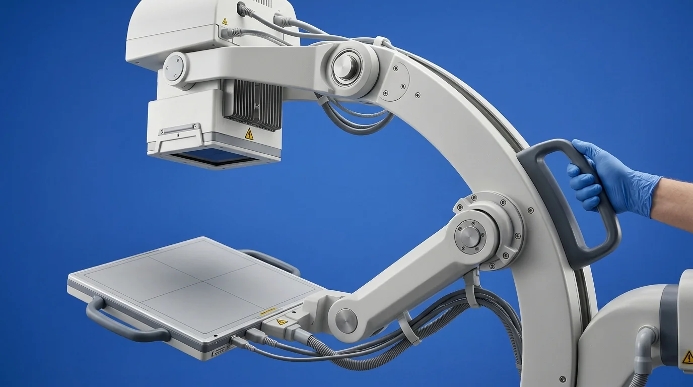 At its heart, a C-arm machine is a sophisticated mobile X-ray system. Its name derives from the characteristic C-shaped gantry that connects an X-ray source and a detector. This design allows for precise, multi-axial rotation around the patient, enabling the clinical team to acquire images from various angles without repositioning.
Key components include:
- X-ray Generator and Tube: This is the source of the X-ray beam. The generator controls the key parameters—kilovolt peak (kVp) and milliampere-seconds (mAs)—which determine the beam's energy and intensity, respectively. Fine control over these settings is essential for optimising image quality while minimising radiation dose.
- The "C" Gantry: The mechanical frame allows for extensive movement, including orbital rotation (angling around the patient), horizontal travel, and vertical lift. The degree of freedom in these movements dictates the clinical versatility of the unit.
- Imaging Detector: II vs. FPD: This is arguably the most critical component, responsible for converting the X-ray image into a viewable format. There are two primary technologies:
- Image Intensifier (II): The conventional technology. An II converts X-rays into light, which is then captured by a camera. While a proven workhorse, II systems can be subject to geometric distortions (like pincushion and S-distortion) and have a larger physical profile.
- Flat Panel Detector (FPD): The current standard in digital imaging. FPDs use a layer of silicon to directly convert X-ray photons into digital signals. This results in superior image quality, a wider dynamic range, no geometric distortion, and a more compact, streamlined design. FPDs also typically offer better performance at lower radiation doses.
- Workstation and Monitor Cart: This is the system's command centre. It houses the computer for image processing, patient data management, and image storage (typically in DICOM format). High-resolution, medical-grade monitors are essential for accurate visualisation.
Key Clinical Applications Across Specialties
The utility of a C-arm machine extends across numerous medical disciplines, each with specific imaging requirements.
- Orthopaedics and Trauma: This is the most common application. A C-arm is essential for visualising fracture reductions, confirming the alignment of bone fragments, and guiding the placement of implants like plates, screws, and intramedullary nails.
- Cardiology and Vascular Surgery: Used for procedures like coronary angiography, angioplasty, and the implantation of pacemakers and defibrillators. A Digital Subtraction Angiography (DSA) feature is critical here for visualising blood vessels.
- Urology: Indispensable for guiding instruments during ureteroscopy, percutaneous nephrolithotomy (PCNL) for kidney stone removal, and placing ureteral stents.
- Pain Management: Provides real-time guidance for targeted injections, such as epidural steroids, nerve root blocks, and facet joint injections, ensuring precise needle placement.
- Neurosurgery: Primarily used in spinal procedures like fusions, discectomies, and vertebroplasty to verify the placement of pedicle screws and other hardware.
- Gastroenterology: A key tool during Endoscopic Retrograde Cholangiopancreatography (ERCP) to visualise the biliary and pancreatic ducts.
Evaluating a C-Arm Machine for Your Facility: A Checklist
Selecting the right C-arm requires a methodical evaluation of its technical specifications against your primary clinical needs.
1. Image Quality and Detector Technology
- Detector Type (FPD vs. II): For new procurements, an FPD is almost always the superior choice due to its better image clarity, lower dose potential, and longevity. However, for facilities with a very specific, limited scope of use, a high-quality II system may be a consideration.
- Detector Size: Common sizes are 9-inch and 12-inch (for II) or 21x21 cm and 30x30 cm (for FPD). A smaller detector is often sufficient for extremities and pain management, while a larger one is necessary for abdominal, vascular, or spinal work.
- Image Processing Software: Evaluate the software's capabilities. Features like edge enhancement, noise reduction, last image hold (LIH), and digital zoom are standard. Advanced functions like DSA or road-mapping are essential for vascular interventions. For facilities requiring a versatile system for procedures ranging from orthopaedics to general fluoroscopy, a machine like the RMS Fluoroscopy C-Arm Machine often represents a balanced configuration, typically featuring a 9-inch Image Intensifier suitable for a wide range of applications.
2. Dose Management and Radiation Safety
- AERB Type Approval: Non-negotiable in India. Ensure the model has valid type approval from the Atomic Energy Regulatory Board (AERB).
- Dose Reduction Features: Look for specific functionalities designed to minimise radiation exposure according to the ALARA (As Low As Reasonably Achievable) principle.
- Pulsed Fluoroscopy: Emits X-rays in short pulses rather than a continuous stream, drastically reducing the total dose.
- Virtual Collimation: Allows the operator to adjust the X-ray field on the last image hold without additional exposure.
- Dose Area Product (DAP) Meter: Measures and displays the radiation dose, which is critical for patient record-keeping and safety monitoring.
3. Operational Ergonomics and Workflow
- Manoeuvrability and Footprint: How easily can the C-arm be moved and positioned in a crowded OT? Check the wheel design, braking system, and overall weight. The unit's footprint must be compatible with your available space.
- Gantry Movement: Assess the range of motion. A large orbital rotation (>115°) and sufficient C-arm depth (isocenter) are needed to perform lateral imaging on larger patients without difficulty.
- Boot-up Time: In an emergency, every second counts. A system that boots up quickly can improve OT turnover and efficiency.
4. Data Management and Connectivity
- DICOM 3.0 Compliance: This is the universal standard for medical imaging. Full DICOM compliance ensures seamless integration with your hospital's Picture Archiving and Communication System (PACS) and Hospital Information System (HIS).
- Storage and Archiving: Check the local storage capacity (number of images) and the available methods for exporting data, such as USB ports or a network connection.
Best Practices for C-Arm Operation and Maintenance
Procuring the right equipment is only half the battle. Proper operational protocols are essential for patient safety, staff protection, and equipment longevity.
- Strict Adherence to Radiation Safety: This is paramount. All personnel present during fluoroscopy must wear lead aprons, thyroid shields, and personnel dosimeters (TLD badges). The principles of time, distance, and shielding must be rigorously applied. The X-ray tube should ideally be positioned under the patient and the detector above to minimise scatter radiation to the operator.
- AERB Compliance and Quality Assurance (QA): Operating a C-arm in India requires strict adherence to AERB regulations. This includes initial registration of the equipment, routine QA testing performed by an AERB-approved agency, and proper record-keeping. These QA tests verify parameters like kVp accuracy, radiation output, and image resolution.
- Preventive Maintenance: Follow the manufacturer's recommended service schedule. Regular preventive maintenance by qualified engineers can prevent unexpected downtime, which is costly in a busy surgical department. Ensuring your C-arm and other critical Medicalequipments are sourced from reputable suppliers with strong service support is a foundational step in maintaining operational readiness.
- Infection Control: The C-arm is often used in a sterile field. It must be cleaned and disinfected between cases according to hospital protocols. Using sterile, disposable drapes designed for C-arms is standard practice.
Equip Your Facility Today
Selecting the right C-arm is a critical decision that impacts clinical capability, operational efficiency, and patient safety. A thorough evaluation of your procedural needs against the technical specifications of available systems is essential for making an informed choice.
For a comprehensive overview of C-arm systems and other diagnostic imaging solutions that meet these demanding clinical and regulatory standards, explore the full range of equipment available on MedikaBazaar.
FAQs

