

Hospitals
Selecting the Right Ophthalmoscope: A Clinical and Operational Guide
A practical guide for clinicians and procurement heads on evaluating ophthalmoscope types, features, and operational considerations for Indian healthcare facilities.


The ophthalmoscope is a cornerstone of physical examination, extending far beyond the ophthalmology department. For clinicians in general medicine, neurology, emergency care, and paediatrics, it is an indispensable tool for assessing systemic health through the unique window of the retina. A clear view of the fundus can reveal the first signs of diabetic retinopathy, hypertensive changes, papilloedema, or embolic events.
For procurement and operations managers, equipping clinical teams with the appropriate ophthalmoscope is not just about a capital asset; it's about enabling diagnostic accuracy, ensuring operational uptime, and standardizing a fundamental examination process across the facility. This guide moves beyond basic specifications to address the practical factors that should inform your selection process, from the technology inside the head to the power source in the handle.
Understanding the Core Technology: Direct vs. PanOptic vs. Indirect
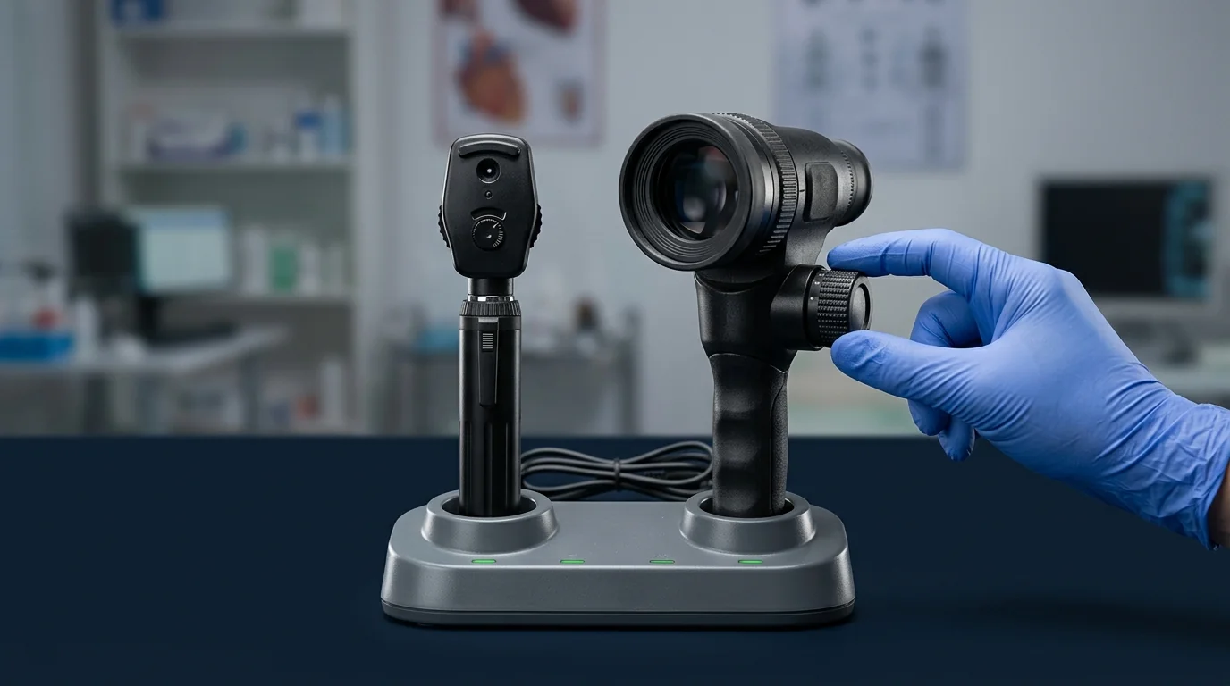 The primary distinction between ophthalmoscopes lies in their optical design, which directly impacts the field of view, magnification, and clinical application.
- Direct Ophthalmoscope: This is the familiar, handheld instrument found in most consultation rooms. It projects a beam of light through the pupil and uses a series of lenses to provide a direct, upright, and highly magnified (around 15x) view of a small segment of the retina. Its narrow field of view (approximately 5-10 degrees) requires skill to navigate the fundus, but it provides excellent detail of the optic disc and macula.
- PanOptic Ophthalmoscope: A significant evolution of the direct ophthalmoscope, this design uses a more advanced optical system to offer a much wider field of view—typically 25 degrees, or five times larger than a standard direct scope. This panoramic view makes it substantially easier and faster to find the optic disc and examine the fundus, particularly through an undilated pupil. This is a considerable advantage in busy OPDs or for non-specialist users.
- Indirect Ophthalmoscope (BIO): Typically a head-mounted unit used with a separate handheld condensing lens (e.g., 20D or 28D), the BIO is a specialist's tool. It provides a stereoscopic (3D), inverted, and wide-field view of the retina. While magnification is lower (2-5x), its true strength is in viewing the entire retinal periphery, which is impossible with a direct scope. It is essential for retinal detachment assessments and in operating theatre settings.
At a glance: a direct comparison highlights the distinct operational roles.
| Feature | Direct Ophthalmoscope | PanOptic Ophthalmoscope | Indirect Ophthalmoscope (BIO) |
| Field of View | Narrow (5-10 degrees) | Wide (25 degrees) | Very Wide (30-50+ degrees) |
| Magnification | High (~15x) | Medium (~5x) | Low (2-5x) |
| Image | Upright, virtual | Upright, virtual | Inverted, real |
| Stereopsis (3D) | No | No | Yes |
| Pupil Dilation | Helpful, not always required | Easier through undilated pupil | Dilation is essential |
| Primary User | GP, Internist, ER, Student | GP, Internist, Paediatrician | Ophthalmologist, Retinal Specialist |
| Primary Use Case | Central fundus, optic disc | General screening, undilated exams | Peripheral retina, surgical settings |
Key Evaluation Criteria for Procurement and Clinical Use
Selecting the right instrument requires looking past the brand name and focusing on the features that impact daily clinical utility and long-term reliability.
Illumination Source: Halogen vs. LED The light source is critical for accurate diagnosis.
- Halogen/Xenon-Halogen: The traditional source, providing a bright, warm light. Many senior clinicians trained with halogen and are accustomed to its colour rendition. However, bulbs have a finite lifespan, generate more heat, and can degrade in colour temperature over time.
- LED: The current standard for new instruments. A high-quality LED source offers a bright, white light with a long operational life (often 20,000+ hours), eliminating the need for bulb replacements. Critically, it provides consistent brightness and colour temperature. When evaluating LED models, look for a high Colour Rendering Index (CRI) to ensure tissue colours appear natural and true.
Power Source: Rechargeable vs. Battery-Operated The choice of handle directly affects operational readiness and recurring expenses.
- Rechargeable Handles: Typically using Lithium-Ion (Li-Ion) or Nickel-Metal Hydride (NiMH) technology, these are ideal for high-volume environments like OPDs and wards. They are stored in a desk or wall-mounted charger, ensuring the instrument is always ready. While the initial investment is higher, it eliminates the recurring need to source and replace disposable batteries.
- Battery-Operated Handles: Using standard AA or C-cell batteries, these handles are lighter and more portable, making them suitable for domiciliary visits, low-volume clinics, or as a reliable backup. The primary drawback is the ongoing need for battery management.
Apertures and Filters A full suite of apertures and filters transforms the ophthalmoscope from a simple viewing device into a versatile diagnostic tool.
- Small Aperture: For viewing the fundus through a small, undilated pupil.
- Large Aperture: The standard setting for general examination with a dilated pupil.
- Slit/Stripe Aperture: Used to assess contours and elevations, such as identifying a swollen optic disc or a retinal lesion.
- Fixation Star/Target: Projects a grid or star for the patient to look at, helping to assess the location and steadiness of fixation.
- Cobalt Blue Filter: Used in conjunction with fluorescein dye instilled in the eye to detect corneal abrasions or ulcers, which will glow green under the blue light.
- Red-Free Filter: Excludes red light to provide high contrast when viewing retinal vasculature and nerve fibre layer defects. Blood vessels appear black, making subtle haemorrhages or neovascularization more apparent.
Diopter Range and Build Quality These factors determine the instrument's focusing capability and longevity.
- Diopter Lens Wheel: This wheel corrects for the refractive error of both the patient and the examiner to bring the fundus into sharp focus. A standard range of approximately -20 to +20 diopters is sufficient for most clinical scenarios. The wheel should rotate smoothly but with distinct clicks for precise control.
- Construction: Instrument heads are typically made from durable polymers or metal. Metal heads offer superior longevity and impact resistance, which is a key consideration for a device used daily in a demanding hospital environment. Check for solid construction and well-sealed optics to prevent dust ingress.
Integrating Ophthalmoscopy into Clinical Workflow
Effective use of the ophthalmoscope depends on its accessibility. A device locked away in a specialist's office is of no use during a medical emergency on the ward. Best practice dictates that an ophthalmoscope should be a standard component of every diagnostic wall unit in consultation rooms and readily available on all emergency crash carts.
Ensuring every examination point is equipped requires a clear procurement strategy. A wide range of diagnostic ophthalmoscopes is available to suit different clinical needs, from basic screening models for general wards to advanced diagnostic sets for specialty departments. Standardizing on a single platform can also simplify staff training and the management of accessories and power sources.
Maintenance, Sterilization, and Compliance
Proper care is essential to protect your investment and ensure patient safety.
- Cleaning and Disinfection: The ophthalmoscope head, particularly the brow rest, should be wiped down between patients using a soft cloth dampened with an approved disinfectant like 70% isopropyl alcohol. Never immerse the instrument head in liquid, as this will damage the optics and internal mechanisms.
- Care of Lenses: The viewing lens should only be cleaned with a lens-safe, lint-free cloth to avoid scratches. Dust can be removed with a soft blower brush.
- Bulb/LED Care: For halogen models, handle replacement bulbs carefully to avoid touching the glass. For LED models, no replacement is typically needed for the life of the instrument.
- Compliance: For procurement, ensure that devices meet recognized quality and safety standards. Look for certifications such as ISO 13485 (for the manufacturer's quality management system) and CE marking, which demonstrates conformity with European health and safety standards. This provides an assurance of manufacturing quality and electrical safety.
Equip Your Facility Today
Selecting the appropriate diagnostic tools is fundamental to high-quality patient care. By systematically evaluating the clinical application, user environment, and essential features of an ophthalmoscope, you ensure your teams are equipped for accurate and efficient examination for years to come.
FAQs

