

Dentist
Integrating a Portable X-Ray Unit into Your Dental Practice: An Operational Guide
An operational guide for clinicians and procurement heads on selecting, integrating, and safely operating a portable x-ray unit in a modern Indian dental practice.


For any modern dental practice, diagnostic imaging is the bedrock of effective treatment planning. While traditional wall-mounted X-ray systems have been the standard, the operational flexibility offered by a high-frequency DC portable x ray unit presents a compelling case for its integration. This is not about replacing established systems wholesale, but about augmenting your diagnostic capabilities, improving workflow, and enhancing patient management, especially in multi-operatory setups or specialised care scenarios.
From a practice management perspective, the decision to incorporate a portable radiographic device goes beyond simple convenience. It impacts chair time, staff movement, patient comfort, and even the physical layout of your clinic. Understanding the core technical specifications, regulatory mandates, and safety protocols is therefore not just advisable—it's essential for a successful and compliant implementation. This guide outlines the key considerations from a peer's perspective, focusing on what truly matters in day-to-day clinical operations.
Core Clinical Applications in Dentistry
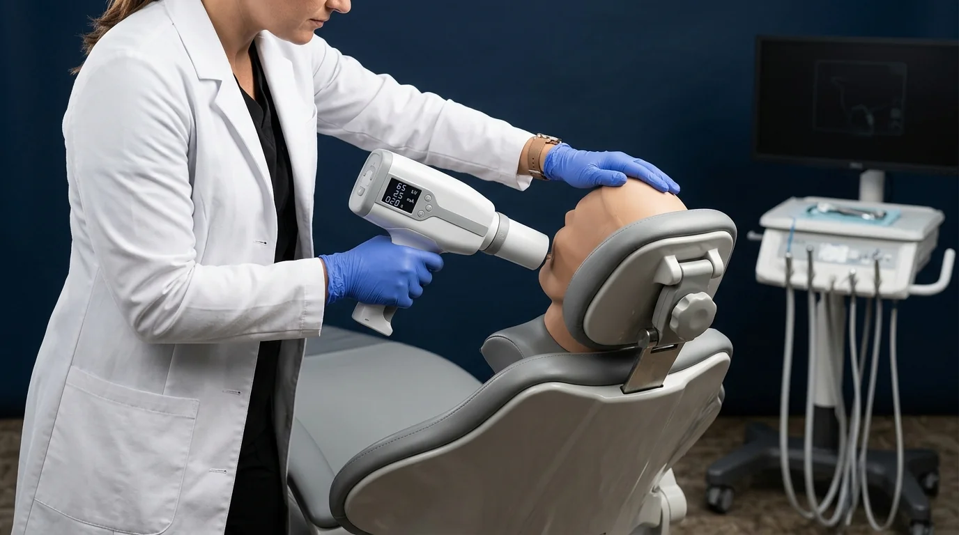 A portable X-ray unit's primary function is to serve as a versatile tool for intraoral radiography. Its mobility allows it to be used effectively across a range of common and specialised dental procedures.
- Standard IOPA and Bitewing Radiography: The most frequent application is for routine periapical and bitewing images. The ability to bring the device to the patient's chair, rather than moving the patient to a dedicated X-ray room, significantly streamlines the process, cutting down on turnaround time between appointments.
- Endodontic Procedures: During root canal treatments, multiple X-rays are often required to assess file position, obturation, and final fill. A handheld unit allows for quick, chairside imaging without requiring the patient to move while a rubber dam is in place.
- Implantology: From pre-operative planning to post-operative verification of implant placement, the ability to take precise, angled shots without repositioning the patient is a distinct advantage.
- Pediatric and Geriatric Patients: For children, anxious patients, or those with limited mobility, a portable device minimises disruption and stress. The imaging process is faster and less intimidating than being moved to a separate room and positioned in front of a large, fixed machine.
- Mobile Dental Camps and Domiciliary Care: For outreach programs or home visits for bedridden patients, a portable unit is indispensable, enabling diagnostic services far beyond the confines of the clinic.
Operational Advantages Over Fixed Units
The decision to specify a portable unit often comes down to clear operational benefits that impact the clinic's bottom line and efficiency.
- Space Utilisation: A new clinic or a renovation of an existing one can save significant space and construction expense by forgoing a dedicated, lead-lined X-ray room. Portable units do not require permanent wall fixtures, freeing up valuable operator-y real estate.
- Workflow Efficiency: In a clinic with three or more dental chairs, a single portable unit can serve all of them. This eliminates patient queues for the X-ray room and reduces non-clinical staff time spent escorting and positioning patients. The entire imaging cycle—from deciding an X-ray is needed to viewing the image on screen—can be completed in minutes at the chairside.
- Flexibility and Scalability: As your practice grows, a portable unit can be easily moved between new operatories without the need for complex installations. It is an asset that adapts to your practice, not one that your practice must adapt to. This flexibility is a key consideration across the entire Dental equipment category when planning for future expansion.
Key Evaluation Criteria for Procurement
When evaluating different portable X-ray models, move beyond the spec sheet and consider how each technical feature translates to clinical performance and operator safety.
Technical Specifications:
- Focal Spot Size: This is a critical determinant of image sharpness. A smaller focal spot (ideally 0.4mm) produces clearer, more detailed images with less geometric distortion, which is vital for accurate diagnosis, especially in endodontics and periodontics.
- Tube Voltage (kVp) and Current (mA): Look for a unit that offers a high tube voltage, typically 60-70 kVp. Higher kVp provides better penetration and produces a greyscale with better contrast, which is essential for differentiating between enamel, dentin, pulp, and bone. The tube current (mA) influences the radiation quantity and exposure time.
- Exposure Time Range: A wide, adjustable range of exposure times allows the operator to fine-tune the settings for different tooth types, patient sizes, and receptor types (RVG sensor vs. phosphor plate), adhering to the ALARA principle.
- Battery Technology: Assess the battery's capacity, typically rated in shots-per-charge. A reliable unit should deliver at least 200-300 exposures on a full charge, sufficient for more than a day of busy operations. Also, consider the charging time and whether the battery is easily replaceable.
Ergonomics and Safety:
- Weight and Balance: The device will be handheld, so its weight and center of gravity are paramount. A well-balanced, lightweight unit reduces operator fatigue and makes it easier to achieve stable, precise positioning for every shot.
- Radiation Shielding: This is non-negotiable. The unit must have integrated backscatter shielding to protect the operator. This usually consists of a lead-impregnated acrylic shield around the collimator. Verify that the design minimises radiation leakage. For a clear example of these integrated safety and performance features, consider a device like the Alerio Neo Portable X-Ray, which is designed with these clinical demands in mind.
Radiation Safety and Regulatory Compliance in India
Operating any radiological equipment in India requires strict adherence to guidelines set by the Atomic Energy Regulatory Board (AERB). For a portable X-ray unit, compliance is focused on both the equipment itself and the protocols for its use.
- AERB Type Approval: The first procurement checkpoint is to ensure the model has valid AERB Type Approval. Operating a non-approved device is a serious regulatory violation. This approval confirms the device meets national standards for radiation safety and performance.
- Operator Protection:
- Correct Technique: The operator must hold the device with their arms extended and keep the backscatter shield parallel to their body. The device should be held stable at the end of the cone.
- Personal Protective Equipment (PPE): Even with shielding, operators must wear a lead apron (0.25mm or 0.5mm lead equivalence) and a Thermoluminescent Dosimeter (TLD) badge to monitor their personal radiation exposure over time.
- Safe Zone: The operator should stand within the designated safe zone, typically behind the plane of the device’s backscatter shield.
- Patient Protection:
- The ALARA (As Low As Reasonably Achievable) principle must always be applied. Use the shortest exposure time necessary to achieve a diagnostic-quality image.
- Patients must be provided with a lead apron and a thyroid collar for every exposure.
- Facility Registration: The dental facility itself must be registered with the AERB, and the installation and use of any X-ray equipment—portable or fixed—must be part of this registration. While a fully lead-lined room may not be mandatory for portable units depending on workload, the walls of the operatory should be of solid construction (e.g., 9-inch brickwork), and the door must be kept closed during exposure. Always consult the latest AERB safety codes for dental diagnostics when planning your setup for both portable and wall-mounted dental x-ray machines.
Choosing the Right Imaging Solution
Selecting a portable X-ray unit is a strategic decision that enhances clinical efficiency and patient care. By focusing on critical specifications like focal spot size, operator ergonomics, and, most importantly, AERB-compliant safety features, you can equip your practice with a tool that provides both diagnostic excellence and operational flexibility.
Evaluate your clinic’s specific needs, operatories, and patient flow to determine how a portable unit can best be integrated. For a comprehensive overview of certified imaging equipment, explore the curated solutions available on MedikaBazaar to find a device that aligns with your practice's standards.
FAQs

