

Dentist
Operational & Clinical Considerations for the Runyes X-Ray and RVG Combo
An operational guide for procurement managers and clinicians evaluating the Runyes portable X-ray and RVG sensor combo for institutional dental use in India.


For any modern dental practice, the transition from conventional film-based radiography to digital is no longer a question of 'if' but 'how'. The decision impacts workflow, diagnostic accuracy, and patient safety. Sourcing these components separately—a dental X-ray source and a digital sensor—can introduce compatibility challenges and procurement complexities. Evaluating an integrated runyes x-ray and rvg combo for clinic presents a streamlined alternative, but requires a thorough assessment of its clinical and operational parameters to ensure it meets your facility’s specific demands.
This resource breaks down the key technical and practical factors to consider when evaluating such a system, focusing on the specifications that directly influence diagnostic output and daily operational efficiency.
Understanding the Core Components: The X-Ray Unit and the RVG Sensor
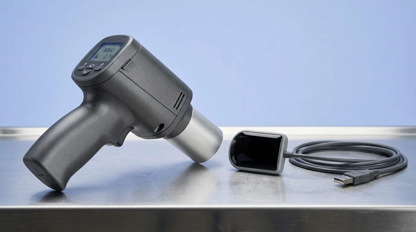 A bundled system comprises two distinct but synergistic pieces of equipment. Understanding each one's role is fundamental to a proper evaluation.
- The X-Ray Unit: The
runyes portable x rayis a handheld, battery-powered intraoral X-ray generator. Unlike traditional wall-mounted units, its primary operational advantage is flexibility. A single unit can serve multiple operatories, eliminating the need for dedicated X-ray installations in every room. This is particularly beneficial for clinics with space constraints or for practices planning future expansion. The core function remains the same: to generate a focused beam of X-radiation for diagnostic imaging. - The RVG Sensor: The Radiovisiography (RVG) sensor is the digital equivalent of radiographic film. It's a solid-state intraoral sensor that captures the X-ray image and instantly transmits it to a computer. This eliminates chemical processing, significantly reduces patient waiting time, and allows for immediate image analysis and enhancement. The
rvg dentaltechnology is central to the efficiency of modern dental radiography.
Key Evaluation Criteria for Institutional Use
When specifying a new dental x ray machine and sensor system, move beyond the brochure and scrutinise the technical specifications that govern performance.
X-Ray Generator Performance
- Focal Spot Size: This is a critical determinant of image sharpness. A smaller focal spot reduces penumbra (blurring at the edges of an object), resulting in a crisper, more detailed image. Look for a focal spot of 0.4mm, which is the standard for high-resolution diagnostic tasks like endodontic canal visualisation or implant thread assessment.
- Tube Voltage (kVp) and Current (mA): These parameters control the energy and quantity of X-rays produced. A higher kVp (e.g., 70 kVp) provides greater penetrating power, which is useful for denser anatomical structures, and creates a longer grayscale (lower contrast). A fixed, optimised kVp and mA, as found in many portable units, simplifies operation while being calibrated for high-quality intraoral imaging.
- Exposure Time: Shorter exposure times minimise the risk of motion artefacts from patient movement and, crucially, reduce the patient's radiation dose. The ability to precisely control exposure time in small increments (e.g., 0.01 seconds) allows for optimisation based on the patient's anatomy and the diagnostic requirement.
RVG Sensor Specifications
- True Resolution (lp/mm): Theoretical resolution can be misleading. The key metric is the true or measurable resolution, expressed in line pairs per millimetre (lp/mm). A higher value (e.g., >20 lp/mm) indicates the sensor's ability to distinguish between two closely spaced objects, which is essential for detecting fine details like early-stage caries or micro-fractures.
- Sensor Technology and Durability: Most modern sensors use CMOS (Complementary Metal-Oxide-Semiconductor) technology, which offers excellent image quality, low power consumption, and high durability. Assess the sensor's construction, particularly the cable connection point, which is a common point of failure. A robust, strain-relieved cable design is an operational necessity.
- Active Area and Ergonomics: Sensors come in standard sizes (e.g., Size 1 and Size 2). Size 1 is often better for paediatric patients or adults with smaller arches, while Size 2 provides a larger field of view for general diagnostics. The physical design, including rounded corners and tapered edges, directly impacts patient comfort and tolerance, leading to better positioning and fewer retakes.
Workflow and Integration
- System Synergy: The primary advantage of a pre-configured combo is guaranteed compatibility. The X-ray generator's exposure settings are inherently matched to the sensitivity of the RVG sensor. Procuring an integrated system like the Runyes Portable X-Ray Machine with Xpect Vision RVG Sensor Combo eliminates the guesswork and potential interoperability issues that can arise when sourcing components from different manufacturers or a general
dental x-ray machine shop. - Software and Connectivity: The imaging software should be intuitive, stable, and DICOM (Digital Imaging and Communications in Medicine) compliant. This ensures images can be easily stored, retrieved, and shared with other systems or specialists. Check for essential tools like measurement, density analysis, contrast enhancement, and annotation.
- Portability and Battery Life: For a
portable dental x ray machine, battery performance is paramount. Evaluate the number of exposures possible on a single charge. A capacity of 300-400 exposures is a strong benchmark, ensuring the device can handle a busy day of clinical work without needing a midday recharge, preventing workflow interruptions.
Radiation Safety and Regulatory Compliance in India
Operating any X-ray equipment in India is governed by the Atomic Energy Regulatory Board (AERB). Adherence is non-negotiable.
- AERB Type Approval: Ensure the specific model of X-ray machine has AERB Type Approval. This is the manufacturer's responsibility and confirms the device meets national safety standards for radiation-generating equipment.
- Facility Level Registration: Your clinic must be registered with the AERB's e-LORA (e-Licensing of Radiation Applications) platform. The approved X-ray unit must be registered to your facility.
- Operator and Patient Safety: Modern portable units incorporate significant safety features, including extensive internal lead shielding to prevent leakage radiation and an external backscatter shield to protect the operator. Even with these features, standard radiation safety protocols, such as maintaining distance and using lead aprons for patients, must be strictly followed. The digital nature of RVG systems supports the ALARA (As Low As Reasonably Achievable) principle by requiring significantly less radiation dose per image compared to D-speed or E-speed film.
Operational Best Practices: From Installation to Daily Use
Proper technique and maintenance are essential for maximising the lifespan and diagnostic value of your imaging system.
- Positioning and Technique: Use beam-aiming and sensor-holding devices (e.g., Rinn holders) to ensure correct parallelism and angulation. This standardises image geometry, prevents cone-cutting, and drastically reduces the need for retakes, saving time and unnecessary patient exposure.
- Sensor Handling and Maintenance: The RVG sensor is a delicate and high-value component.
- NEVER autoclave the sensor. Heat and pressure will destroy it.
- Always use a new disposable barrier sheath for each patient to prevent cross-contamination.
- Clean the sensor between uses with a manufacturer-approved, non-alcoholic disinfectant wipe. Alcohol can damage the sensor housing over time.
- Avoid sharp bends or tension on the cable, especially at the sensor connection point.
- Image Archiving: Implement a robust, routine backup protocol for all digital patient images. This is a clinical and legal requirement. A combination of local and cloud-based storage is advisable to protect against data loss.
Equip Your Facility Today
Selecting the right diagnostic imaging system is a foundational decision for any dental practice. By focusing on the core technical specifications, workflow integration, and regulatory requirements, you can specify a solution that enhances diagnostic confidence and streamlines clinical operations.
For your institutional requirements, explore the detailed specifications of integrated dental imaging solutions on MedikaBazaar to find the right fit for your clinical workflow.
FAQs

