Complete Cautery Machine Guide


Cautery Machine Fundamentals
Electrosurgical units are used in endoscopic procedures to cut tissue and control bleeding by delivering controlled electrical energy. Most systems are designed to operate in both monopolar and bipolar modes, which differ mainly in how the electrical circuit is formed and completed.
Monopolar vs Bipolar Modes
In monopolar cautery machine, electrical energy is delivered to the target tissue through an active electrode, where the intended tissue effect occurs. The current then travels through the patient’s body and exits via a dispersive electrode (return pad) placed on the skin, completing the circuit back to the generator.
In bipolar electrosurgery, the electrical current is confined to the treatment area. Both the delivery and return of energy occur through the same instrument, such as a bipolar forceps or probe, eliminating the need for a dispersive electrode.
The choice between monopolar and bipolar techniques depends on clinical requirements. Monopolar mode is commonly used for its versatility, while bipolar mode is often preferred in patients with implanted electronic devices, such as pacemakers, to minimize the risk of electrical interference.
Power Settings by Procedure
| Model Category | Typical Product Example | Power Range | Operating Modes | User Interface | Common Applications |
| Entry-level Electrosurgical Unit | Standard Electrosurgical Cautery | Low to medium | Monopolar cutting and coagulation | Basic analog controls | Dressing areas, minor procedures, training use |
| Compact Clinic Model | Mini Cautery Mono / Dual-Mode System | Up to ~100 W | Monopolar with focused dual-electrode | Analog or basic digital | ENT clinics, dermatology, small operating rooms |
| Analog OT System | Analog Cautery System (250W / 400W) | 250–400 W | Monopolar with focused dual-electrode | Rotary dials and meters | General surgery, gynecology, orthopedic OTs |
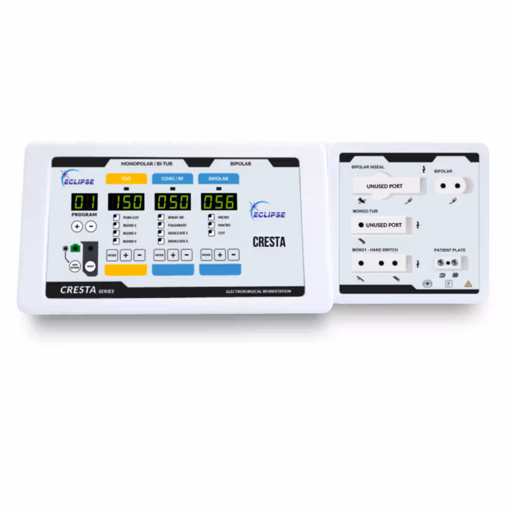
| Digital OT Cautery | Digital Cautery System (300W / 400W) | 300–400 W | Full monopolar with focused dual-electrode options | Digital display and controls | High-throughput, multi-specialty theatres |
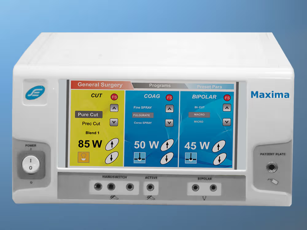
| Advanced Digital ESU | BIOHEARTZ 400W (480 kHz) | 400 W | Advanced monopolar with focused dual-electrode | Precision digital interface | Corporate hospitals, teaching institutions, complex procedures |
Electrode Types & Selection
Electrocautery electrodes are typically manufactured from stainless steel or tungsten and are classified based on their mode of energy delivery monopolar or bipolar. Each electrode type is designed with specific tip shapes to suit different surgical requirements.
Monopolar Electrodes (Active Electrodes)
Needle / Wire Electrodes
Designed for high-precision cutting and localized coagulation. Commonly used where fine control and minimal tissue disruption are required.
Blade / Spade Electrodes
Versatile electrodes suitable for both cutting and coagulation in routine surgical procedures.
Ball Electrodes
Used for surface coagulation and fulguration, particularly effective for controlling superficial bleeding and treating broad lesions.
Loop Electrodes (LEEP)
Primarily used for tissue excision and lesion removal, especially in gynecological and dermatological procedures.
Bipolar Electrodes
Bipolar Forceps
Allow current to flow only between the two tips of the instrument, offering precise coagulation with minimal impact on surrounding tissues. Ideal for procedures involving nerves and delicate vessels.
Spatula / Tweezer Tips
Used for grasping tissue while simultaneously achieving coagulation, offering controlled handling during surgery.
Patient Safety Protocols
Safety Precautions for Using Electrosurgical Devices
When setting up an operating room or procedure area for electrosurgical equipment, following basic safety practices is essential to protect both patients and staff.
Ensure proper training
Safe operation depends largely on the user. Physicians and assisting staff should be adequately trained, confident in handling the device, and familiar with the manufacturer’s instructions and precautions.
Use smoke evacuation
Electrosurgical procedures generate surgical smoke that may contain harmful substances. Installing an effective smoke evacuation system helps reduce exposure and maintains a safer operating environment.
Use equipment as intended
Always operate devices and accessories strictly according to their specified purpose. Use the lowest effective power settings and avoid substituting or modifying accessories, as improper use increases the risk of injury.
Check equipment before use
Electrosurgical units should undergo routine inspection and testing. Never use a device on a patient unless it has been properly checked, adjusted if needed, and confirmed to be functioning correctly.
Keep flammable materials away
Electrosurgery can create sparks that may ignite flammable substances. Ensure alcohol-based prep solutions, flammable anesthetics, and oxygen-rich environments are carefully managed and kept at a safe distance.
Exercise caution with implanted devices
Patients with pacemakers or other electronic implants require special attention, as electrosurgical currents may interfere with device function. Consultation with specialists is recommended before proceeding.
Prevent contact with metal objects
Metal contact can increase the risk of burns. Remove jewellery from patients and staff, and check instruments to ensure there are no exposed metal surfaces during the procedure.
Avoid skin-to-skin contact
Unintended skin contact along the electrical pathway can lead to alternate site burns. Proper patient positioning and correct placement of the return electrode help reduce this risk.
Handle active electrodes carefully
When not in use, place the electrode in a designated insulated holder. Avoid bringing the active tip too close to tissue unintentionally, and remain attentive throughout the procedure to prevent accidental injury.
Routine Maintenance Schedule
After each use, cautery tips should be promptly placed in a 0.5% chlorine decontamination solution for about 10 minutes to reduce bioburden. This solution can be prepared using household liquid bleach or bleach powder. Following decontamination, clean the tip using a soft-bristled brush and a mild detergent solution, brushing lengthwise to protect the wire. Continue cleaning until all visible organic material is removed.
Once cleaned, soak the cautery tip again in a fresh 0.5% chlorine solution for a minimum of 20 minutes. After soaking, rinse thoroughly with boiled and cooled water to eliminate any residual chlorine. Allow the tip to air dry completely, then either use it immediately or store it in a dry, covered container until the next procedure.
Before starting a surgical procedure, place a sterile sleeve over the handpiece, insert the cleaned tip, and briefly activate the unit to confirm that the tip heats evenly. This step helps verify proper device function and provides surface sterilization.
Alternative high-level disinfection methods, such as chemical disinfectants or steam sterilization, may also be used when appropriate. However, repeated autoclaving may affect plastic components of the tip and should be done with caution.
OT Setup Optimization
Optimizing the operating theatre setup is essential for ensuring procedural efficiency, safety, and smooth workflow. Proper placement of equipment, clear cable management, and easy access to essential instruments help reduce delays and minimize the risk of contamination or accidental injury. Organizing devices based on frequency of use, maintaining adequate spacing around the operating table, and ensuring reliable power and ventilation support uninterrupted procedures and enhance overall surgical performance.
Related Products

Fastest by 14th Apr
COD
Basco Digital Surgical Diathermy
₹71,250
₹92,42923% OFF
₹70,250 with MBFIRST
Fastest by 13th Apr
COD
Delcatt Micro-controlled ESU unit
₹77,700
₹93,24017% OFF
₹76,700 with MBFIRST
Fastest by 14th Apr
COD
Gemi Micro Computer Controlled (GEM 300) ESU unit
₹75,000
₹79,9206% OFF
₹74,000 with MBFIRST
Fastest by 14th Apr
COD
Gemi Shortwave cum Pulse 1000W ESU unit
₹49,950
₹59,94017% OFF
₹48,950 with MBFIRST
Fastest by 14th Apr
COD
Gemi Micro Computer Controlled ESU unit
₹43,750
₹46,6206% OFF
₹42,750 with MBFIRST


FAQs
SOCIAL MEDIA
DOWNLOAD APP
HELP US IMPROVE
SOCIAL MEDIA
DOWNLOAD APP